Elbow osteochondritis dissecans (OCD)
What is elbow osteochondritis dissecans (OCD)?

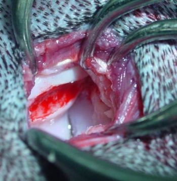
Humeral OCD lesion
Osteochondrosis is a developmental condition that arises due to a disturbance in the normal differentiation of cartilage cells resulting in failure of endochondral ossification (an essential process during development of the skeletal system resulting in bone formation). In dogs that grow very quickly, the rapid cartilage growth can outstrip its own blood supply causing abnormal cartilage development resulting in lameness, pain and subsequent osteoarthritis. In some cases, flaps of diseased cartilage become separated from the remaining cartilage surface. This is called osteochondritis dissecans (OCD).
Is there a particular breed predisposition?
Genetic factors are the most important cause of OCD with strong breed predispositions, particularly in Labradors and giant breed dogs. Different breeds appear to be predisposed to developing the condition in different joints. Various other factors such as dietary or nutritional problems during the first few months of life, hormonal imbalances and joint trauma are also thought to increase the risk of developing OCD.
How do I know if my dog has OCD?
Most dogs with OCD start to show clinical signs before they are one year old, although occasionally (particularly with shoulder OCD), signs may present when your dog is older. The clinical signs are variable and depend on the joint affected and the size of the cartilage defect. The most common signs include lameness, stiffness, joint pain or swelling, reluctance to exercise or play, or general depression.
How is elbow OCD diagnosed?
OCD is typically diagnosed following a multimodal evaluation process. Firstly, your dog will be examined by one of our orthopaedic clinicians. Following this, your dog will be admitted to the hospital to allow radiographs of the affected joints under sedation or general anaesthesia. Because OCD can occur at the same time as other developmental orthopaedic diseases (such as certain manifestations of elbow dysplasia), some dogs may require additional diagnostic imaging such as CT or MRI which will be performed by our advanced diagnostic imaging team. Your dog will receive one-to-one nursing care throughout the process by one of our nurses from the prep nursing team who are all highly trained and experienced in anaesthesia and sedation. Following diagnostic imaging, your dog may require surgery to allow arthroscopy (keyhole surgery) to be performed on the affected joint to allow a close-up examination inside the joint.
Will my dog develop osteoarthritis?
As soon as OCD starts to develop, osteoarthritis (inflammation of the joint and associated bones) immediately starts to develop. Once present, osteoarthritis cannot be cured but can be effectively managed in most patients.
How is elbow OCD treated?
Treatment of elbow OCD depends on the size of the cartilage defect. In small defects, debridement of the loose flap may be chosen. Large defects are usually treated by resurfacing. Fitzpatrick Referrals have published several papers on OCD treatment and have pioneered the use of osteochondral autograft transfer (OAT) in canine patients. Building on this work, we have been pivotal in the development of the synthetic osteochondral replacement (SOR) and the most recent advancement in the field of osteochondral repair; the SynACART™ implant.
Your orthopaedic clinician will advise you on the best course of treatment for your dog.
Synthetic osteochondral resurfacing (SOR)
Synthetic osteochondral resurfacing (SOR) involves implantation of a synthetic implant, made from a medical-grade polycarbonate plastic, to fill the defect caused by the OCD lesion thus restoring the surface of the articular cartilage.
SynACART™
Manufactured by Arthrex®, SynACART™ represents the cutting edge of treatment for canine OCD. The implant is a second-generation synthetic osteochondral resurfacing implant which incorporates a smooth, biocompatible polycarbonate urethane surface to reduce friction and combines this with a trabecular titanium metal allowing bone to grow into the implant.

