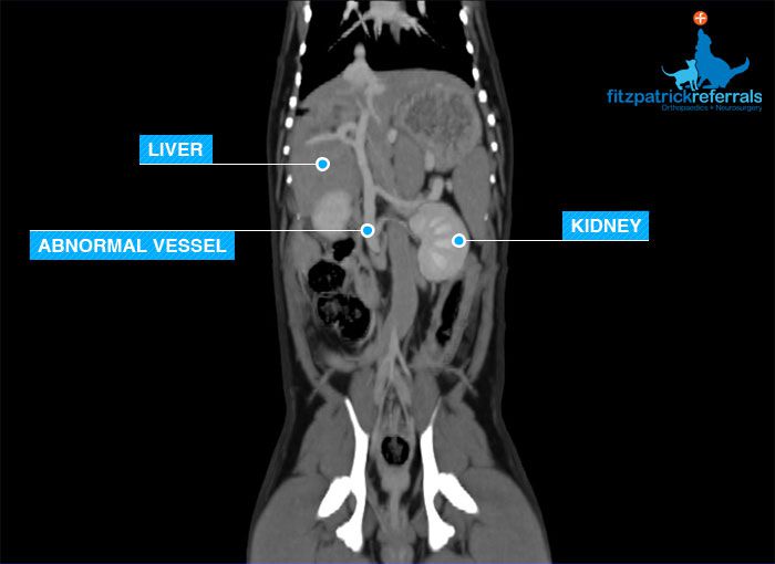
Olly, a one year old Pug, was referred to us with a history of recently having difficulties in passing urine. His primary care vet had taken a urine sample and run some blood tests, and had identified that he also had a liver problem. It was found that Olly had bladder stones, but as he was still quite a young dog, our concern was that these stones may be of a type that can form as a result of disturbed metabolism encountered when a dog is born with a congenital abnormality of the blood supply to the liver (a portosytemic shunt).
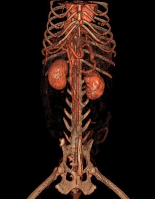
The CT scanner has the ability to process the data it collates during a scan and reconstruct this into 3-dimensional images. 3-D images help the surgeon to identify structures with greater ease and increased accuracy. In this patient the reconstruction allowed the easy identification of the rogue vessel which can be extremely difficult amongst the abdominal viscera.