On 8th November we celebrate World Radiography Day – the anniversary of when x-rays were first discovered. Since that all-important day in 1895, x-rays have found an integral role in both human and animal healthcare and here at Fitzpatrick Referrals, they are used on a daily basis as a crucial diagnostic tool for many of our patients.
We’re looking back at Monty’s story and the important role imaging played during the course of his specialist treatment with us.
Monty’s story
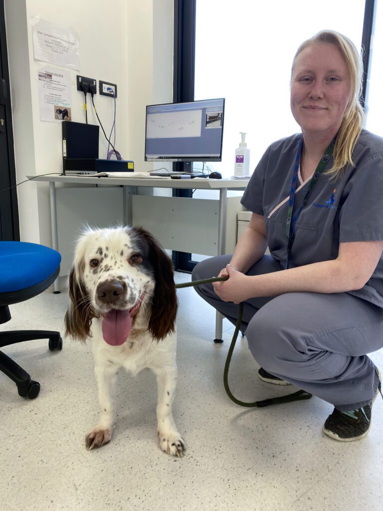
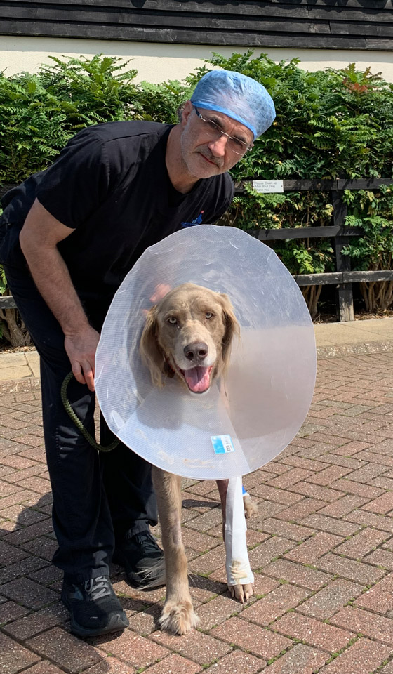
English Springer Spaniel Monty came to see us when he was 11 months old after struggling with lameness and noticeable deformities on both his forelimbs.
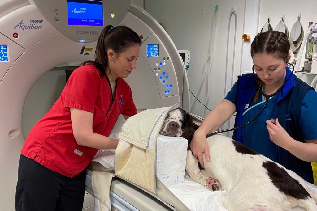
Orthopaedic Registrar Mario Coppola sent Monty for a CT (Computed Tomography) scan and radiographs to further investigate what was happening with Monty’s bones and the soft tissue surrounding them.
During a CT scan, x-rays are continuously emitted while rotating 360 degrees around the body, acquiring high-resolution three-dimensional data sets that are sensitive to tiny bony changes and can be reconstructed in many planes.
After reviewing Monty’s CT, Mario diagnosed him with bilateral antebrachial growth deformity (ABGD) – an angular and/or rotational deformity of the forelimb. After a discussion with Monty’s family, together it was decided that surgery to correct the deformity was the best option for Monty.
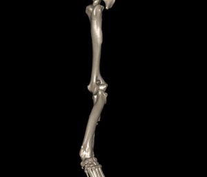
The CT data was used by our bioengineers at Fitzbionics to design the 3D printed cutting guides and two custom implants – one for each of Monty’s front legs and designed to fit exactly to every curve and contour of Monty’s bones.
3D reconstruction of Monty’s right forelimb rendered from the CT data pre and postoperatively
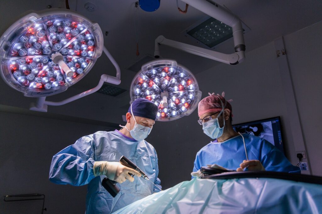
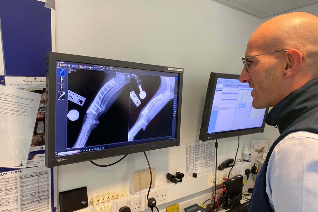
Monty underwent his surgery and immediate postoperative plain film x-ray imaging was performed to check the position of the implants. The x-ray images are two dimensional, quick to obtain and exhibit excellent contrast between bony anatomy, metalwork and the overlying soft tissue.
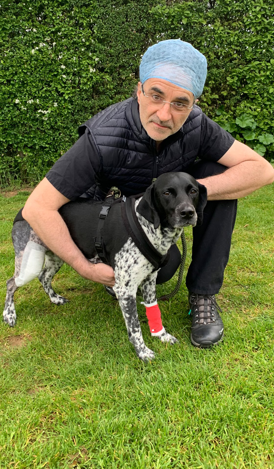
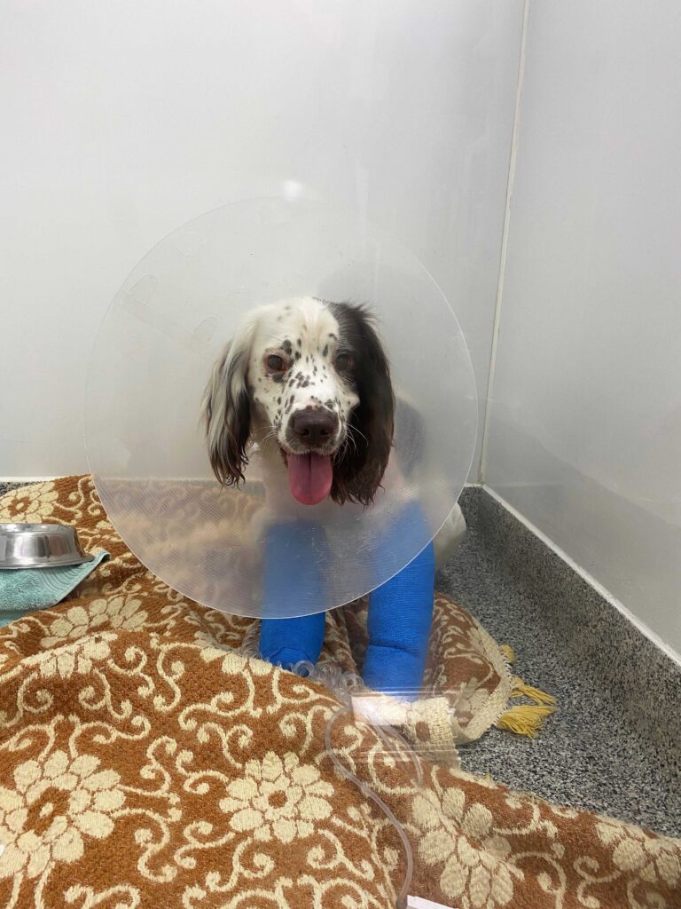
After careful review, Mario was satisfied with the placement of the implants and the new alignment of Monty’s forelimbs and Monty was taken to the ward area to recover. Monty ended up staying with us a little while longer as he developed some sores on his paw but ultimately made good progress.
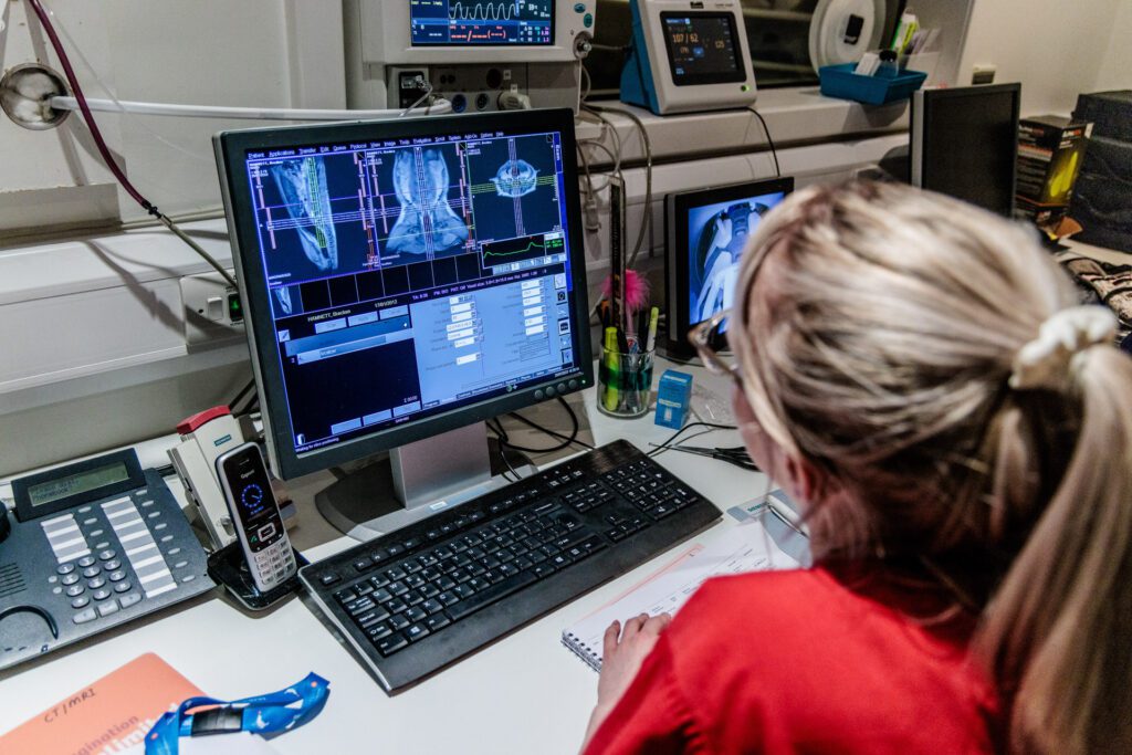
He came back to see Mario for 6 and 12-week check-ups where further CT and x-rays were carried out to ensure the implants were still anchored neatly into the bone and the bones themselves were healing well.
Monty’s x-ray images of his right forelimb
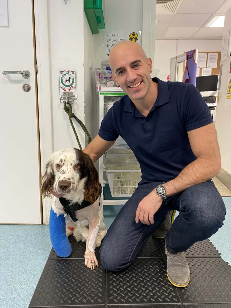
Monty’s recovery
Monty had his final recheck appointment five months after surgery and we were so pleased to hear from his family that he no longer had any lameness, was gradually building up his exercise levels and back to enjoying life!