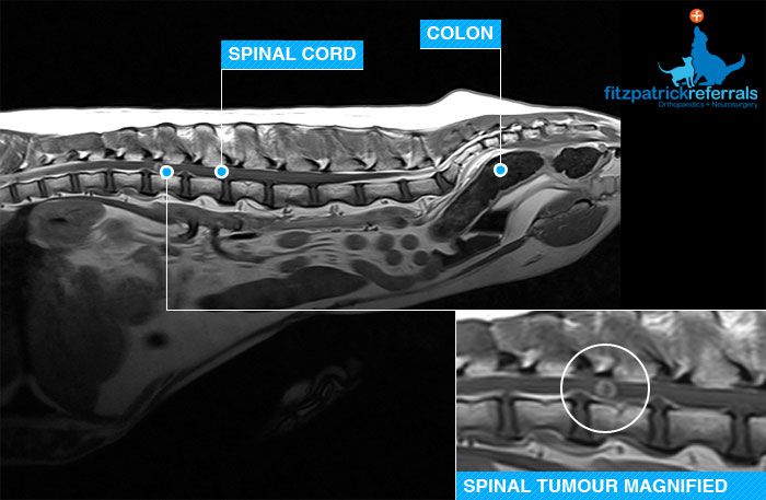
The above image is an MRI of the thoracolumbar spine of an eleven-year old Hungarian Visla. The dog presented to the primary care vet with a history of recent onset hind limb lameness. Although both limbs were affected the left hind appeared worse than the right and the owners found he was unable to jump into the car. Exercise restriction and anti-inflammatory therapy had been tried but after an initial improvement the dog relapsed.
Due to the relapse the patient was referred to the outpatient imaging service for an MRI examination of the spine. A marble sized mass was identified just in-front of the first lumbar vertebra. There was a high index of suspicion that this mass represented a neoplasm (cancer) and so a sample of cerebrospinal fluid, the fluid that bathes the spinal cord, was sent away for analysis. Sadly in this case the lesion was confirmed as neoplastic and the patient was sadly euthanized after a couple of happy weeks at home with her owner.
Sometimes the information gained from advanced imaging gives the answers that allow an end of life decision to be made for a patient. It makes sure all avenues of treatment are explored and ensures the right ethical decision is made for the patient.