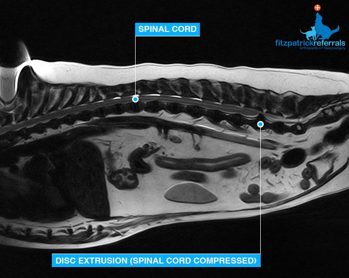
This 9 year old crossbreed was referred to the outpatient diagnostic imaging service for the investigation of spinal stiffness. Owners had noticed that over the past year or so she was progressively more reluctant to climb the stairs or jump on the sofa. Recently they had noticed she had a hunched back, she was shivering and shaking, not weight bearing on her left hindleg and collapsing on her right. Above is an image from her MRI study showing very obvious compression of the spinal cord in the region of the fifth and sixth lumber disc space. Loss of nerve sensation and function is quite common with this type of lesion which can lead to incoordination, weakness, paralysis and incontinence. Removal of the offending disc material from the spinal canal relieves the pressure on the cord. This delicate procedure is called a hemilaminectomy. This dog was operated on by the referring vet and to date is doing well highlighting the importance of gaining a proper insight into the underlying pathology causing common clinical symptoms.
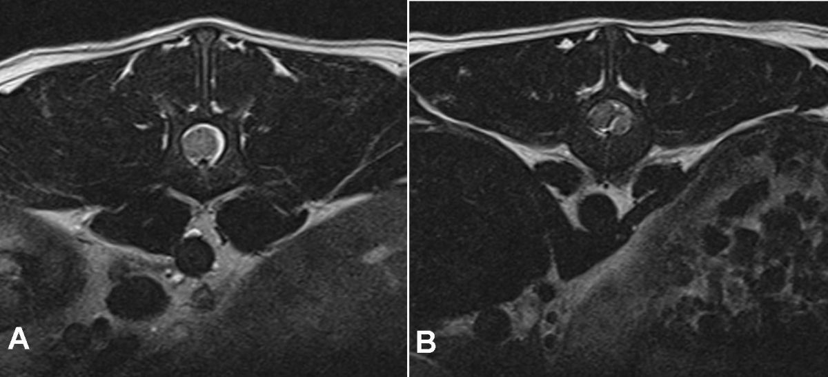
A and B above are crossectional views of an MRI scan demonstrating what a normal non-compressed spinal cord looks like and then in B you can see compression of the spinal cord by disc material. Disc lesions can occur in the cervical, thoracic or lumbar regions. Spinal conditions can be treated medically or surgically. Nonsurgical (medical or conservative) treatment is recommended for animals with apparent pain only or animals that have mild neurologic deficits but are able to walk. Usually restricted exercise and anti-inflammatory therapy are tried and the response to treatment is closely monitored. Surgical treatment involves spinal surgery consisting of a ventral slot, hemilaminectomy, dorsal laminectomy, disk fenestration etc. Animals with disc lesions causing the loss of deep pain sensation require urgent surgical attention as such extensive compression of the spinal cord can result in irreversible spinal cord damage and permanent paralysis.