
8th November is World Radiography Day and marks the day when x-rays were first discovered in 1895.
Since then, x-rays have gone on to become an integral and indispensable diagnostic tool in modern-day medicine and have given rise to all sorts of innovative imaging modalities, including CT (computed tomography), MRI (magnetic resonance imaging) and ultrasound.
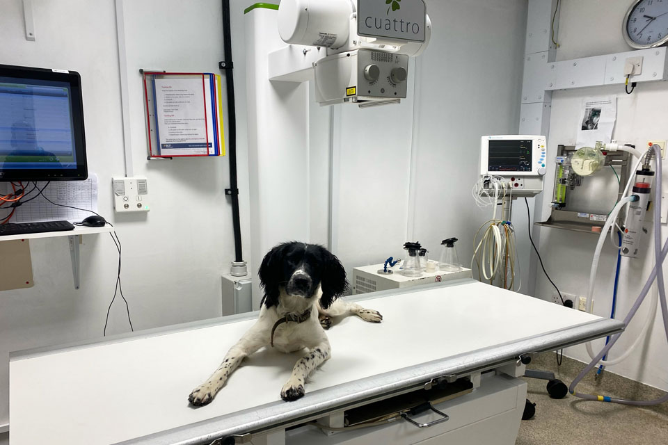
Here at Fitzpatrick Referrals, the large majority of our patients will require at least one of these imaging techniques to diagnose their condition and subsequently ensure that they receive the most appropriate and effective treatment.
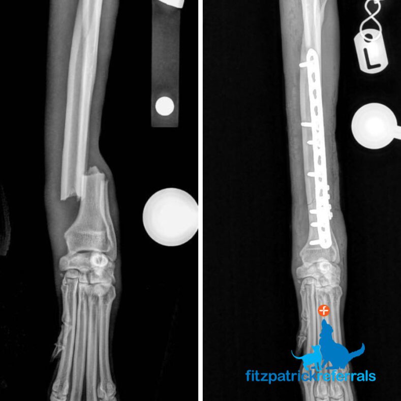
X-ray
Krispy the whippet came to us after suffering a trauma to his left forelimb. X-rays revealed a fracture at the left distal radius and ulna which was fixed using a plate and screws. X-rays were also taken postoperatively to check the position of the metalwork and to ensure the broken bones were back in alignment.
MRI
We saw Sidney the dachshund as an emergency after he had suddenly lost the use of his back legs. An urgent MRI was done of the spine to see if there was anything impinging the spinal cord which would inhibit the neurological signals from Sidney’s brain reaching his hind limbs. The MRI showed a disc extrusion at the level of the L3/L4 vertebrae with disc material in the spinal canal. Sidney was taken straight to theatre so the disc material could be removed and the spine decompressed.
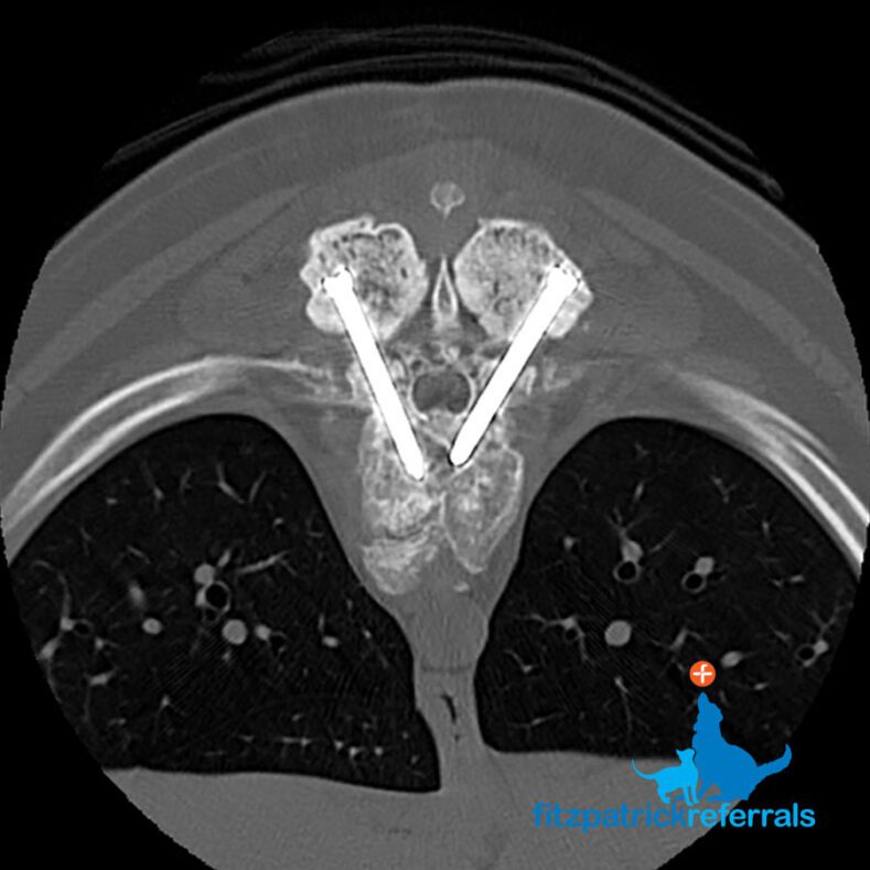
CT
Lottie the boxer came in with ongoing spinal pain and discomfort. Initial investigations and imaging diagnosed Lottie with a degenerative spinal condition which had led to malformations of the bony vertebrae in the thoracic spine. Lottie underwent surgery to relieve pressure on her spinal cord and the spine between T7 and T11 was re-aligned and stabilised using pins and cement. A postoperative CT was performed to look at the spinal column in cross-section and check the pins were anchored into the bone of the vertebral bodies and not in the spinal cord itself.
Wishing everyone (in particular all radiology departments, both human and veterinary) a very happy World Radiography Day.
Read more about our Advanced Diagnostic Imaging Service