What is vestibular disease?
The vestibular system is responsible for maintaining balance, posture and the body’s orientation in relation to the horizon. The vestibular system is comprised of a receptor, located close to the middle ear, a short nerve and the lower stem of the brain.
Vestibular disease is one of the most common neurological presentations in veterinary neurology and can be one of the most challenging. The vestibular system or apparatus, as it is also known, is responsible for maintaining balance, posture, and the body’s orientation in space. This system also regulates locomotion and other movements and keeps objects in visual focus as the body moves. The vestibular system is comprised of the vestibular apparatus itself, the vestibulocochlear nerve, and those parts of the brain that interpret and respond to information derived from these structures. Vestibular disease has been described as a ‘sudden non-progressive, disturbance in balance’.
The commonest underlying cause of vestibular disease is a sudden onset problem that affects one half of the system i.e. one ear. It is a disease more commonly found in older dogs and therefore may also be referred to as ‘old-dog vestibular syndrome’ or ‘canine idiopathic vestibular syndrome’ but is also found to occur in cats in rare instances.
The vestibular system comprises the vestibule and three semicircular canals of the inner ear. Like a carpenter’s spirit level, these structures work with the brain to sense, maintain, and regain balance and a sense of where the body and its parts are positioned in space.
How can I tell if my dog has vestibular disease?
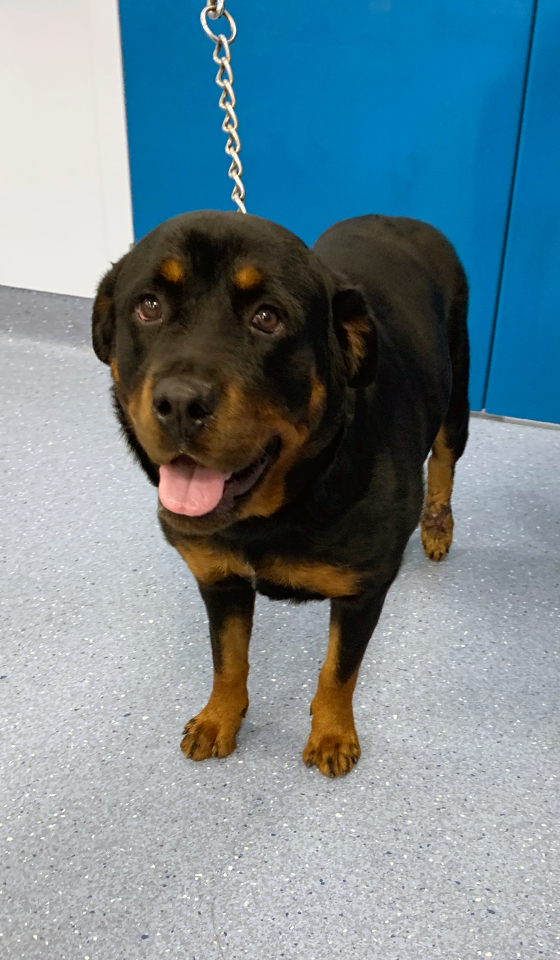
Dogs with vestibular disease usually present with a loss of balance, disorientation, head tilt and irregular jerking eye movements known as nystagmus. Depending on the severity and localisation of the vestibular disease many, but not all dogs, will also be reluctant to or unable to stand or walk and may struggle to do so which can be quite distressing for the dog but also you, the owner. Most dogs will lean or fall in the direction of their head tilt. Damage to the vestibular system can result in some, but not necessarily all of the following clinical signs:
- Asymmetric ataxia i.e. drunken gait
- Abnormal posture e.g. leaning or head tilt (towards the side of the problem)
- Circling or deviating (towards the side of the problem)
- Wide-based stance
- Nystagmus (rapid eye flick)
- Vestibular (positional) strabismus i.e. a squint, typically in a downward direction when the head position is changed
- Vomiting usually due to motion sickness / dizziness
What is the cause of vestibular disease?
We typically split vestibular disease into one of two ‘syndromes’ (collections of clinical signs) on the basis of whether we suspect the condition to involve the peripheral components (ear and nerve) or the central components (parts of the hind brain).
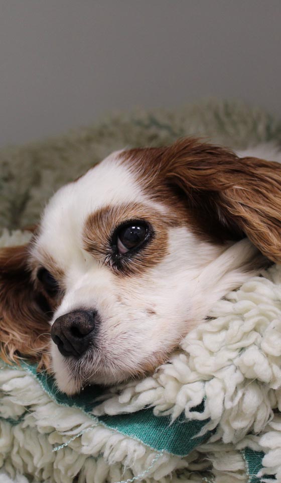
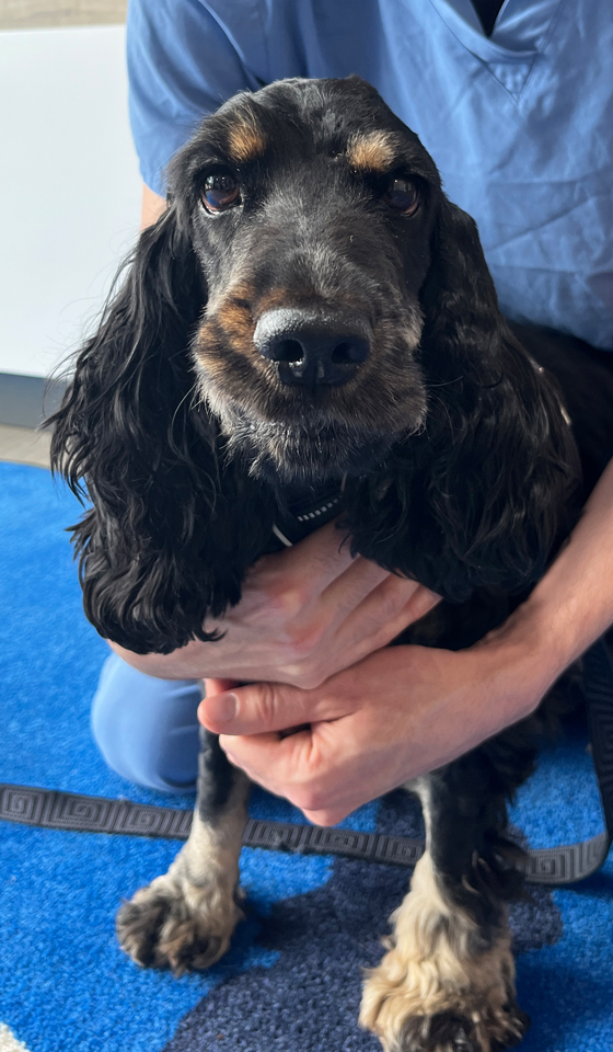
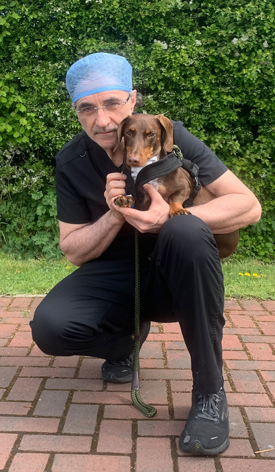
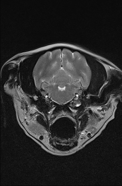
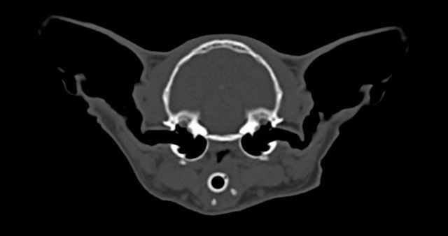
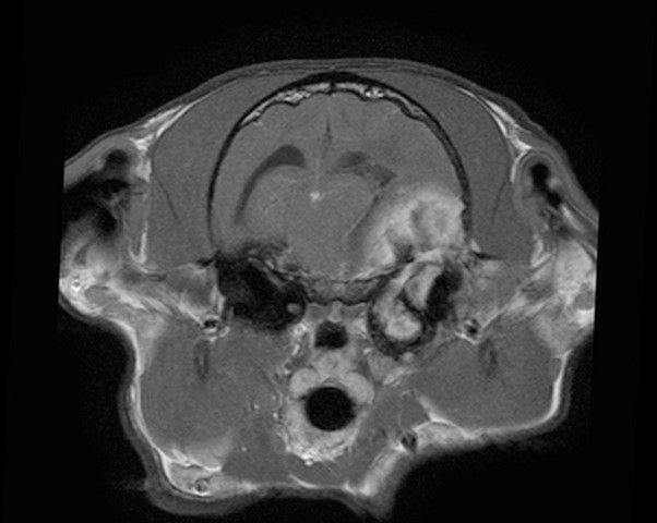
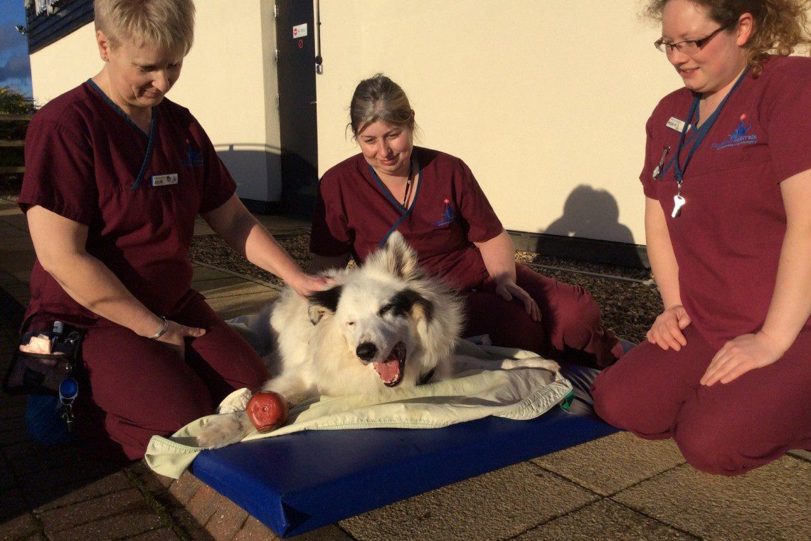
For treatment and prognostic purposes it is vital to locate the source of the problem and distinguish between peripheral vestibular and central vestibular disease. The most reliable indication of central vestibular disease is depressed mental status (e.g. poorly interactive and disorientated) and postural deficits i.e. loss of strength and proprioception (i.e. the sense of where the limbs are in space). Disease causes of vestibular disease this can be divided into various components to include; metabolic, neoplastic, inflammatory, nasopharyngeal polyps, toxica or trauma to name but a few. Your neurology clinician will be able to advise you of the likely source causing your dog or cat to have vestibular disease. The following images illustrate how vestibular disease may present itself:
How is vestibular disease diagnosed?
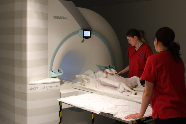
Following a sudden onset of vestibular clinical signs, vestibular disease may be strongly suspected. However, the most important ‘test’ is a thorough neurological examination as this is used to determine if the vestibular syndrome is peripheral or central and will indicate the necessary pathway to further investigation and treatment required. However diagnostic imaging is usually required to confirm a diagnosis. The most useful is MRI as this allows both the ears and the brain to be assessed. However, ear infections can also be identified on CT and in some cases with radiographs.
We have MRI and CT scanners on-site managed by our advanced diagnostic imaging team, which allows rapid diagnosis and differentiation between peripheral and central vestibular disease. MRI scan is the most advanced imaging technique and provides more information than radiographs for diagnosis, however, patients must lie completely still for their MRI scans, and this is only possible with the use of general anaesthesia. CT may only require very profound sedation to achieve optimal stillness and positioning. Your pet will have dedicated one-to-one care during their MRI or CT scan by one of our nurses from the prep nursing team who are all trained and experienced in anaesthesia.
Additional tests which may be recommended are cerebrospinal fluid (CSF) analysis in which the fluid bathing the brain and spinal cord is analysed for abnormalities in protein concentration, cell counts and other parameters. This test is most useful to determine if there is an inflammatory and / or infectious disease process causing your pet’s clinical signs. Tests for infectious agents such as viruses and protozoa may also be appropriate which involves the collection of a blood sample or urine. To determine if your pet has an inflammatory disease process causing clinical signs, a sample of fluid from your pet’s inner ear may be collected during a procedure called myringotomy. Myringotomy involves the insertion of a blunt-ended needle (sprools needle) through the tympanic membrane (eardrum) whereby the neurologist is then able to aspirate a sample of the mucus that is normally produced around the inner ear apparatus. In patients with inner ear infections, this may appear as a pus-like fluid. The collection of this fluid is important as the neurologist can request a bacteriology culture to be performed and with the results, be able to prescribe the most appropriate medication to treat the inflammation. Some neurologists may also use Brainstem Auditory Evoked Response (BAER) to assess the hearing pathways which are close to the vestibular pathways. Deafness, either temporary or permanent is another factor to identify alongside all other clinical signs.
If diagnostic tests are not possible e.g. for financial reasons then the best course of action is to monitor your pet’s neurological status by performing regular and thorough neurological examination. Furthermore, knowing if your pet’s disease is central (brain) or peripheral (ear or nerve) can be a guide as to both prognosis and management.
How is vestibular disease treated?
Vestibular disease is predominantly a non-surgical condition but some underlying causes of the disease may require a degree of surgical intervention. Either way, management vestibular disease requires a twofold treatment pathway:
Dealing with the underlying cause (if possible)
Management of peripheral vestibular disease underlying conditions can vary from medical intervention such as antibiotics or adjunctive surgical or radiotherapy treatment for example in neoplastic disease of the middle or inner ear whereby complete surgical resection of tumours can be difficult or has been unsuccessful. However, cats diagnosed with nasopharyngeal polyps usually require surgical intervention to remove the polyps.
Management of central vestibular disease tends to involve medical management with antibiotics, corticosteroids, antifungal or antiepileptic medication. The specific underlying cause of the disease will determine which treatment is best for your pet.
Providing supportive patient care
Physiotherapy of vestibular disease and supportive therapy are the most important considerations in the rehabilitation of vestibular disease (peripheral or central). During your pet’s hospitalisation period, supportive nursing care will be provided by a multidisciplinary team of neurology clinicians, patient care team, chartered physiotherapists and hydrotherapists. Your dog will be assessed by our specialist team of chartered physiotherapists and they will design and begin a physiotherapy and rehabilitation programme specifically for your dog to help them get back on their feet. Physiotherapy plays a vital part in the treatment of patients with vestibular disease since inactivity and recumbency results in decreased joint movement, stiffness and muscle weakness and contracture which all hinder a smooth road to recovery.
What is the prognosis of vestibular disease?
The prognosis for recovery from vestibular disease is very much dependent on the underlying cause of the clinical signs. The rate and extent of recovery is variable and difficult to predict but can take days, to weeks or months and some patients may have some residual deficits e.g. a subtle head tilt.
Your pet may require treatment for a week or so, or maybe months to ensure the underlying cause of the vestibular disease is managed well. Whereas some of the central vestibular diseases may require life-long treatment. Your neurologist will be able to give you a more accurate prognosis for your pet once they have performed a thorough neurological exam and have a made a definitive diagnosis of the cause of your pet’s clinical signs.