This World Radiography Day, we’re celebrating the vital role that diagnostic imaging plays in the care of our patients here at Fitzpatrick Referrals.
From X-rays and dental radiography to advanced CT and MRI scans, these technologies enable our team to look beneath the surface — helping us diagnose conditions accurately, plan treatments, and improve outcomes for the animals entrusted to us.
World Radiography Day is on 8th November each year, to remember the anniversary of when X-rays were first discovered in 1895 by German physicist Wilhelm Röntgen.
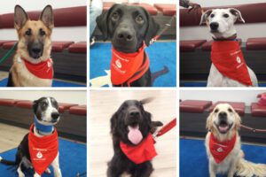
To mark the occasion, we’re highlighting the incredible work of our imaging team and showcasing the capabilities of our diagnostic suite. We’ve enlisted a few of our colleagues and their own much-loved dogs who have received specialist treatment with us, to share the role that imaging played in their particular treatment journey.
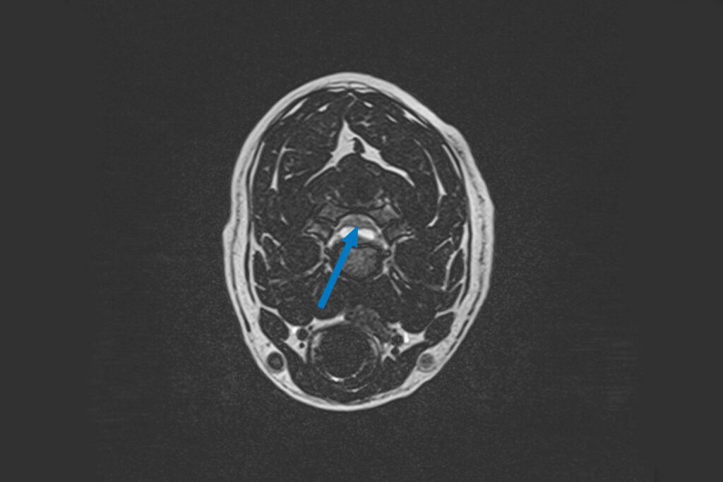
Bryn’s MRI for severe tetraparesis (loss of mobility in all limbs)
(Our 1.5T MRI scanner generates a high magnetic field and a series of radio waves to map out water molecules within body)
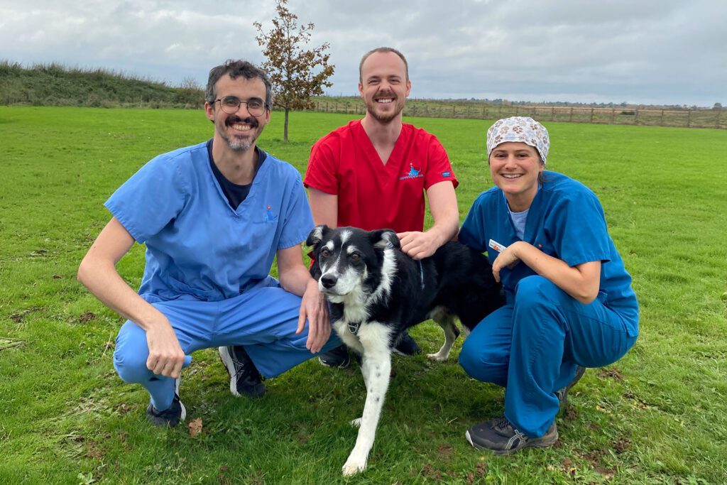
When Senior RVN Sam’s beautiful border collie Bryn suddenly lost the use of all four of his limbs he was referred as an emergency to see Neurologist Marc Pérez Soteras. A problem in Bryn’s neck was quickly suspected and he was rushed in for a MRI scan of that area.
Our 1.5T MRI produces excellent contrast between various soft tissue structures in any plane of the body and is the gold standard for spinal imaging. In this instance, it was needed to visualise all the discs in Bryn’s neck, his spinal cord and nerve roots to identify what exactly was stopping the signals from his brain reaching his legs and where that was located. MRI revealed a hydrated disc extrusion at the level of C3-C4, which was compressing the spinal cord and starting to affect the nerves that control his breathing.
He was taken into surgery, where Marc was able to successfully decompress the disc and relieve pressure on the spine, restoring the use of his legs, allowing him to resume his daily walks with Sam and his siblings.
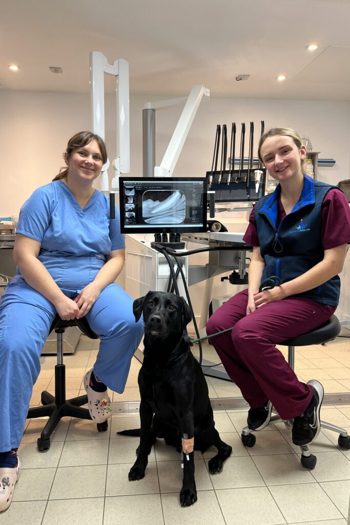
Max’s dental radiographs for his fractured mandibular canine tooth
Radiography Assistant Ellie brought her lovely Labrador Max in to see dentist Hannah after he fractured his lower canine tooth.
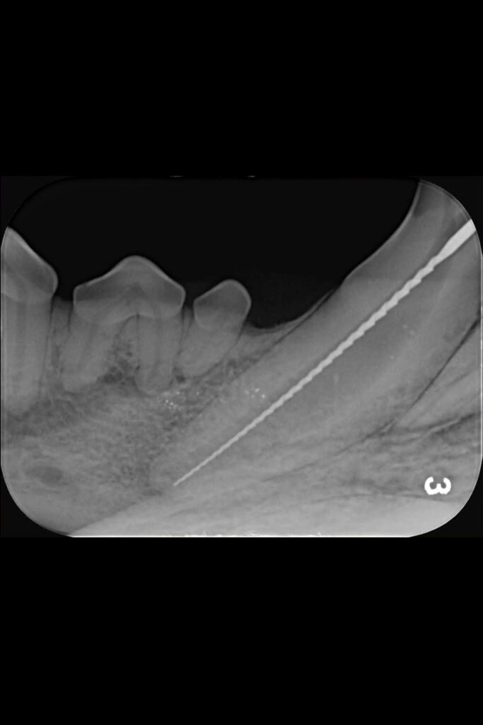
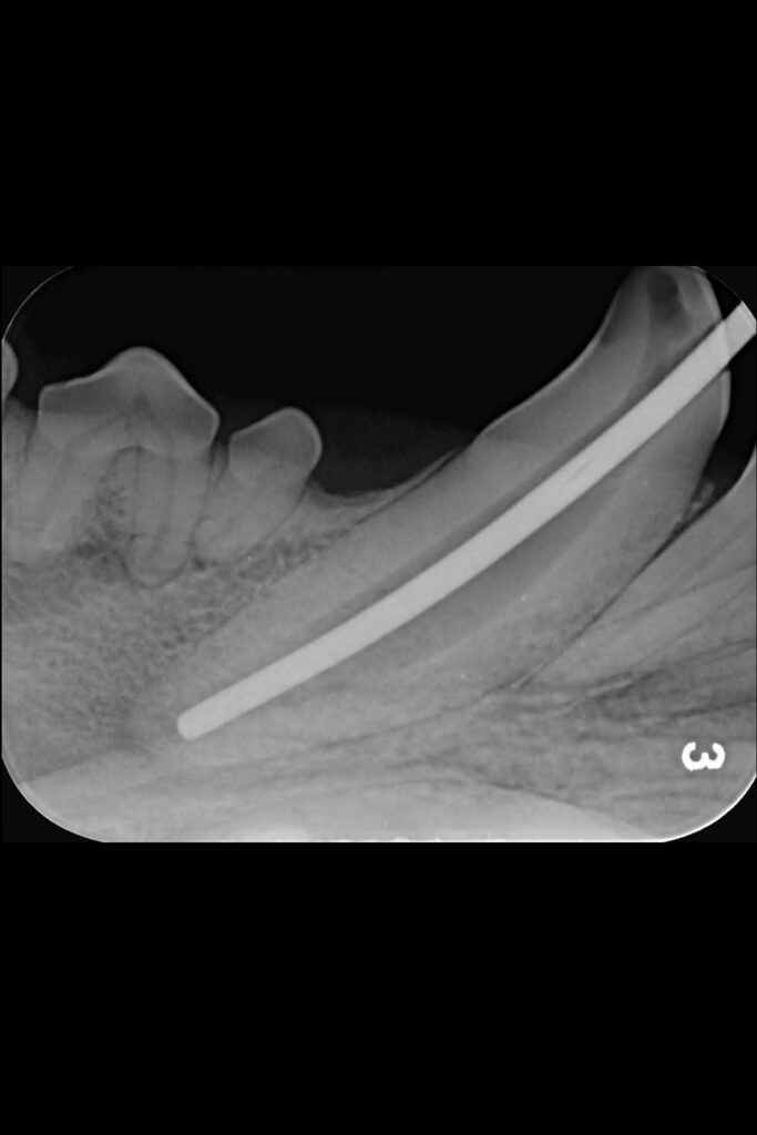
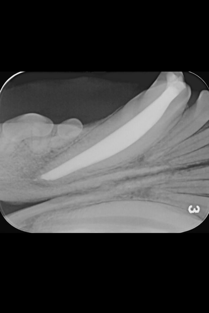
Initial radiographs were taken on our intraoral dental-specific X-ray unit to evaluate the dense tooth and bone structure. This revealed what was happening within the tooth and beneath the gumline – all the areas that cannot be seen with a visual examination. These showed not only the crown fracture but also a wide pulp chamber, which required root canal treatment.
A key part of this treatment involves inserting increasing file sizes into the tooth to reach the pulp to remove any infected or necrosed tissue. X-ray images are then again taken at various points throughout the procedure to check file size and position.
Max was a great boy for his procedure and is now back to working hard as a gundog.

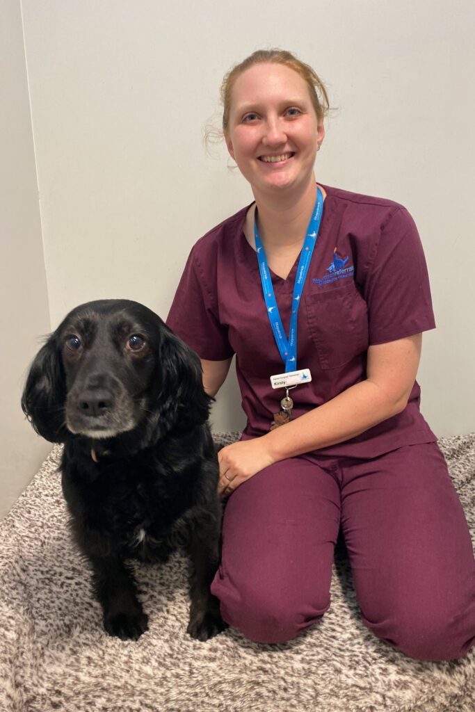
Rosey’s X-rays for her hip dysplasia
Super spaniel Rosey came in with her mum, Senior Surgical Technician Kirsty, with some ongoing lameness and stiffness on her hind limbs.
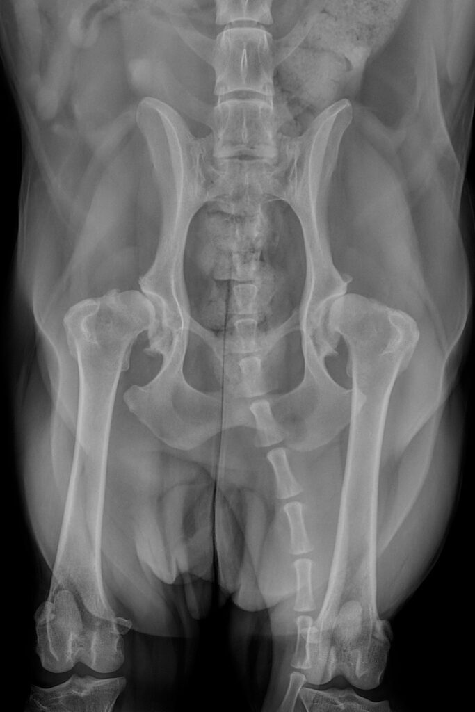
As a first step, her orthopaedic surgeon requested for some radiographs to be taken using one of our three X-ray units to better understand what was happening in Rosey’s bones and joints. X-ray images are really effective at showing bony detail and providing an overall assessment of the bone integrity and alignment.
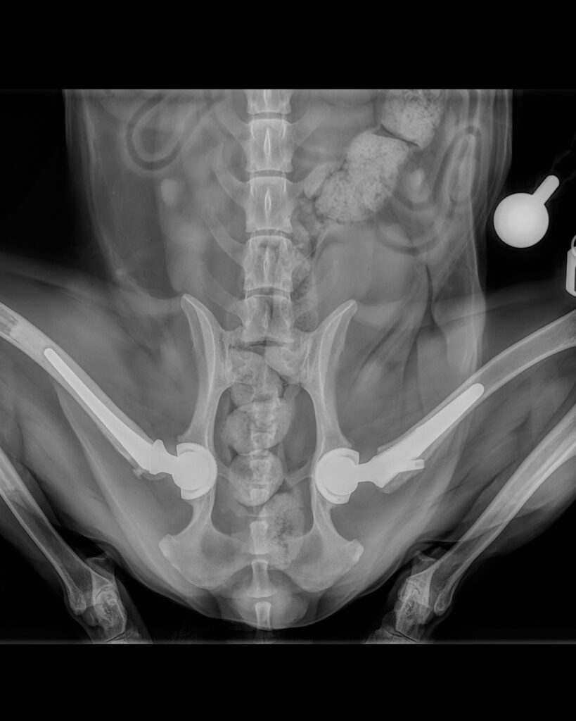
They showed that poor Rosey was suffering from bilateral hip dysplasia and associated osteoarthritis, but fortunately, total hip replacements were a treatment option and Kirsty opted to go ahead with the procedure for her canine friend, replacing the right hip first and the left nine months later.
Postoperative radiographs were also taken immediately following surgery and at regular intervals after each operation to demonstrate good implant placement and bone healing.
Rosey made a swift recovery and is now zooming around on her new hips, enjoying life pain-free.
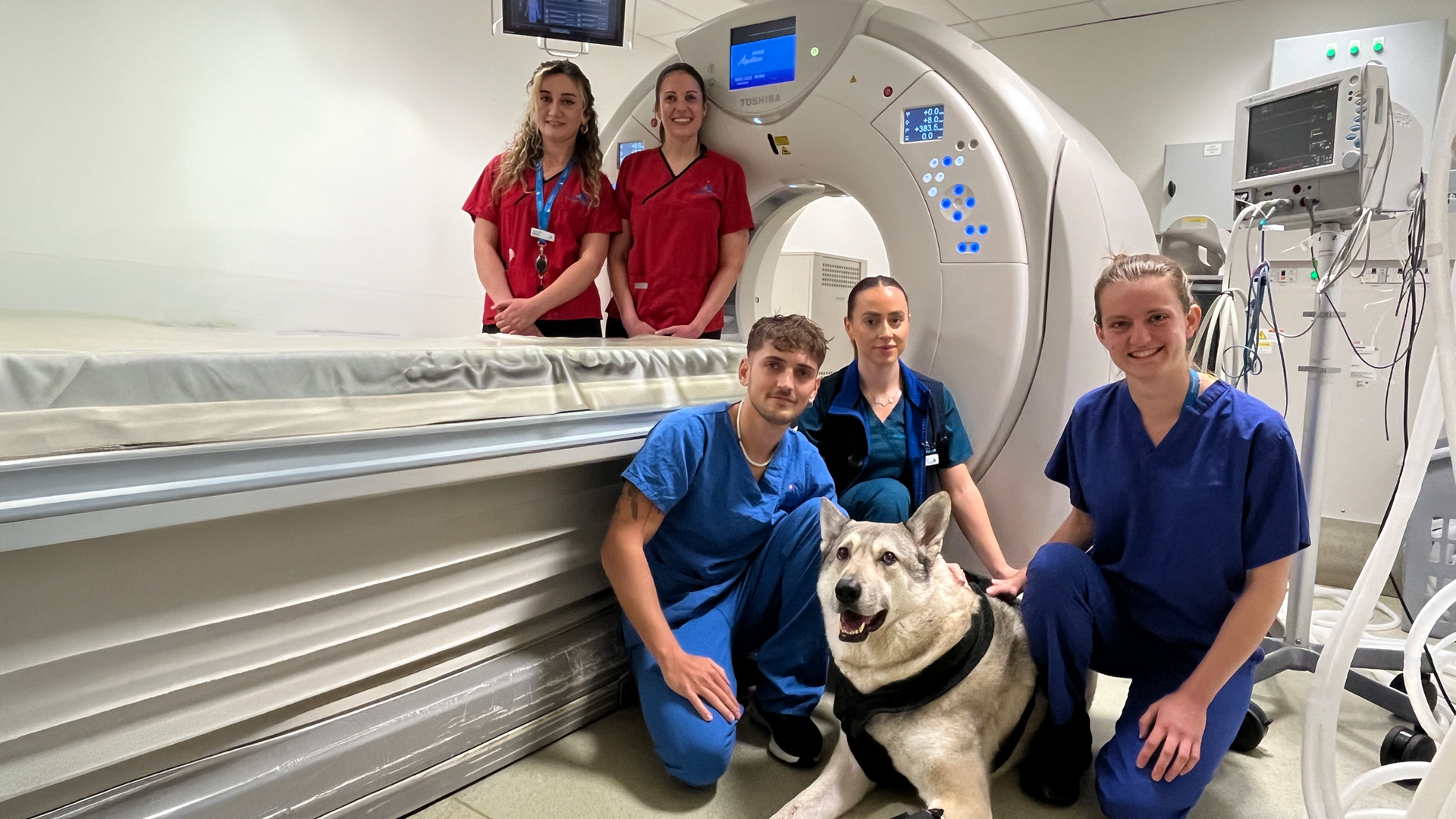
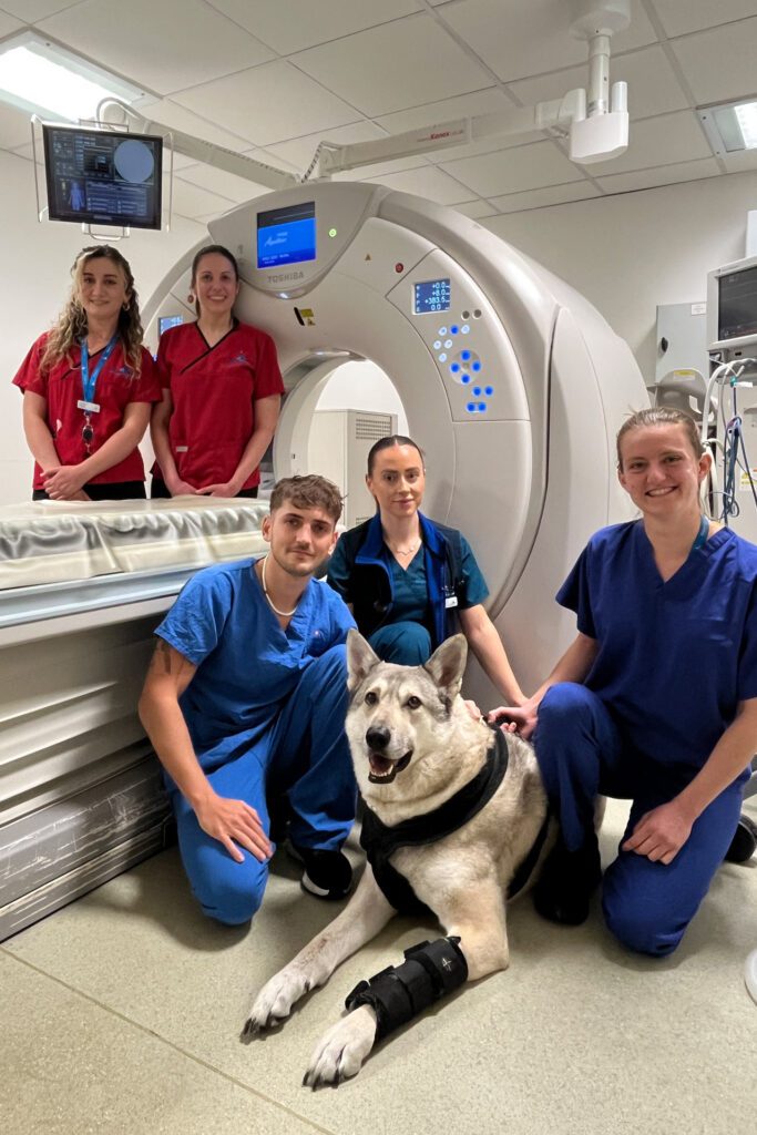
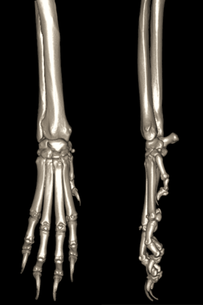
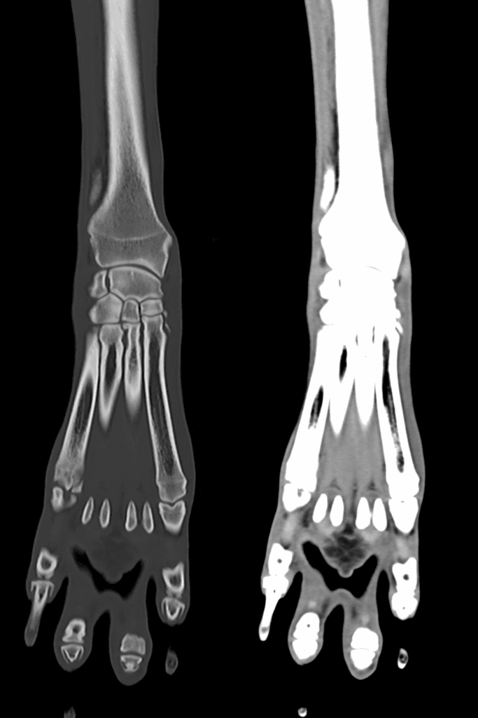
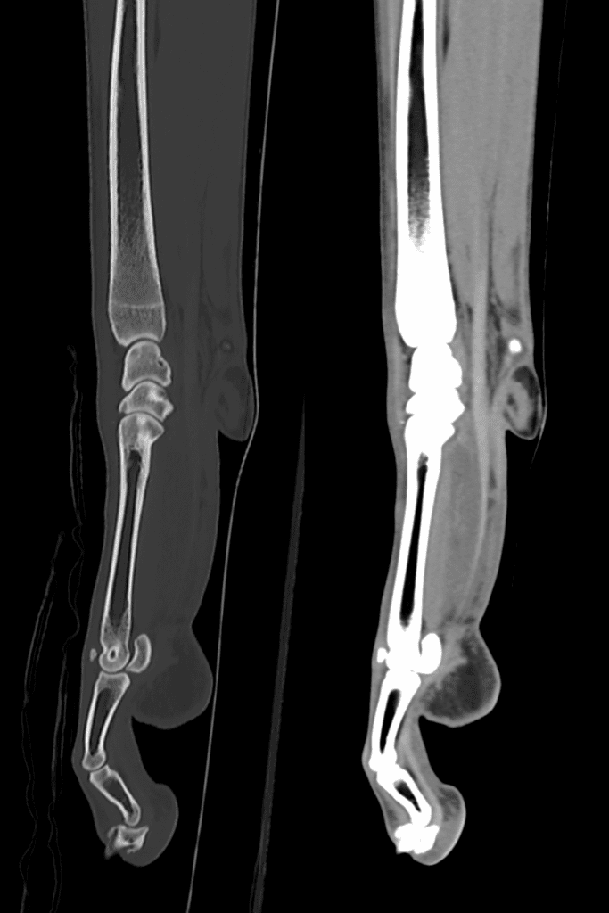
Nico’s CT scan for his carpal (wrist) laxity
Nico the wonderful wolfhound was struggling to weight bear on his right carpus and his neurologist mum Nadia Shihab brought him to see our orthopaedic team.
A physical examination showed carpal laxity on the right carpus so his orthopaedic surgeon immediately referred Nico for a CT scan using our 160-slice CT scanner to help him work out exactly what was happening in that joint. The high-resolution CT images were obtained in seconds, offering superior resolution of the bony anatomy, identifying small and subtle pathology and allowing 3D reconstruction of Nico’s carpus.
The diagnosis was progressive carpal collapse and treatment options included a pan carpal arthrodesis using a custom implant designed and modelled from the CT images so that it could fit Nico’s bones perfectly.
Nico had his surgery and is now recovering, getting stronger day by day with Nadia by his side every step of the way.
Wishing all radiology departments (both human and veterinary) a very happy World Radiography Day!
Read more about our Advanced Diagnostic Imaging service