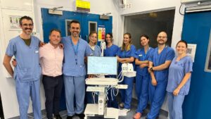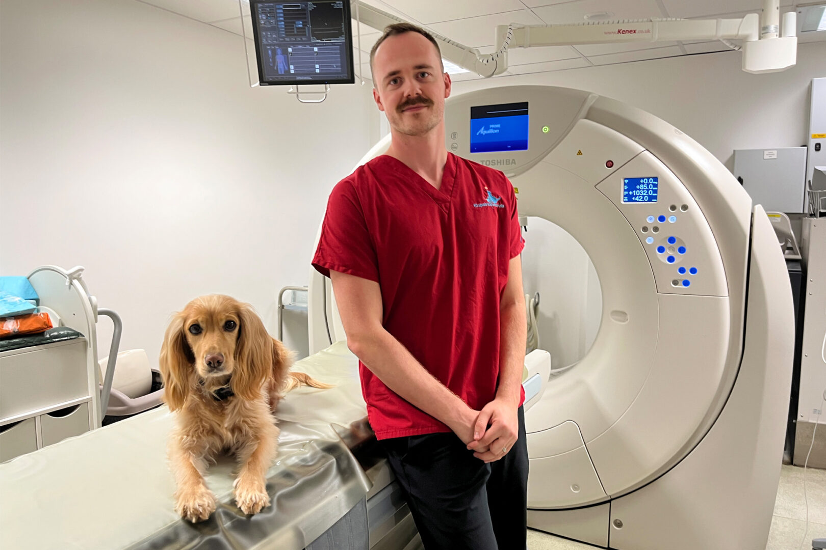
In November, we celebrate World Radiography Day and our Advanced Diagnostic Imaging team. This year, we highlighted the crucial role veterinary diagnostic imaging plays in our patients’ treatment journeys.
Every day brings different families, different questions, and different images…but one shared purpose: to provide the best possible care for our patients.
A discovery that changed medicine
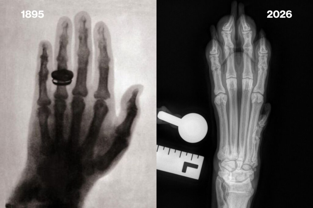
X-rays were first discovered in November 1895 by German physicist Wilhelm Röntgen, while he was experimenting with something else entirely.
What began as an unexpected discovery transformed medicine forever, allowing us to see inside the body in ways that had never been possible before. This laid the foundations for the medical imaging technology we know and use today.
Radiographs – seeing the unseen
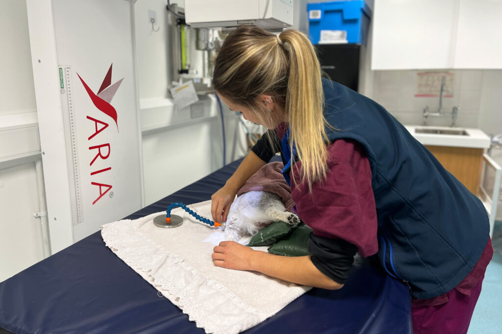
Radiographs are quick, efficient and incredibly valuable and are often one of our first steps in investigating a patient’s symptoms. They can assess the bones and joints, help identify fractures and other traumatic injuries and look for changes associated with conditions such as osteoarthritis.
A simple image can provide some very important answers.
Some common examinations:
- Pelvis and hindlimb radiographs
- Elbow radiographs
- Chest radiographs
Some of the questions they can help us investigate:
- Does the patient have a cruciate rupture?
- Can we identify features of hip dysplasia or osteoarthritis?
- Are we dealing with a fracture or a dislocation or both?
CT – looking deeper
CT (computed tomography) uses rotating X-rays to capture the body in 3D, allowing us to view anatomy in cross-section. When we need a closer look, CT is there to separate out structures and identify smaller or more subtle changes – it is especially good at demonstrating the bones and the lungs.
More detail + more angles + more planes = a clearer picture.
Some common examinations:
- CT of the elbows and forelimbs
- CT of the pelvis to toes
- CT of the chest and abdomen
- Some of the questions it can help us investigate:
- Are there signs of developmental elbow disease and elbow osteoarthritis?
- Can we assess limb alignment in 3D?
- Is there evidence of a tumour or cancer spread?
MRI – a different view
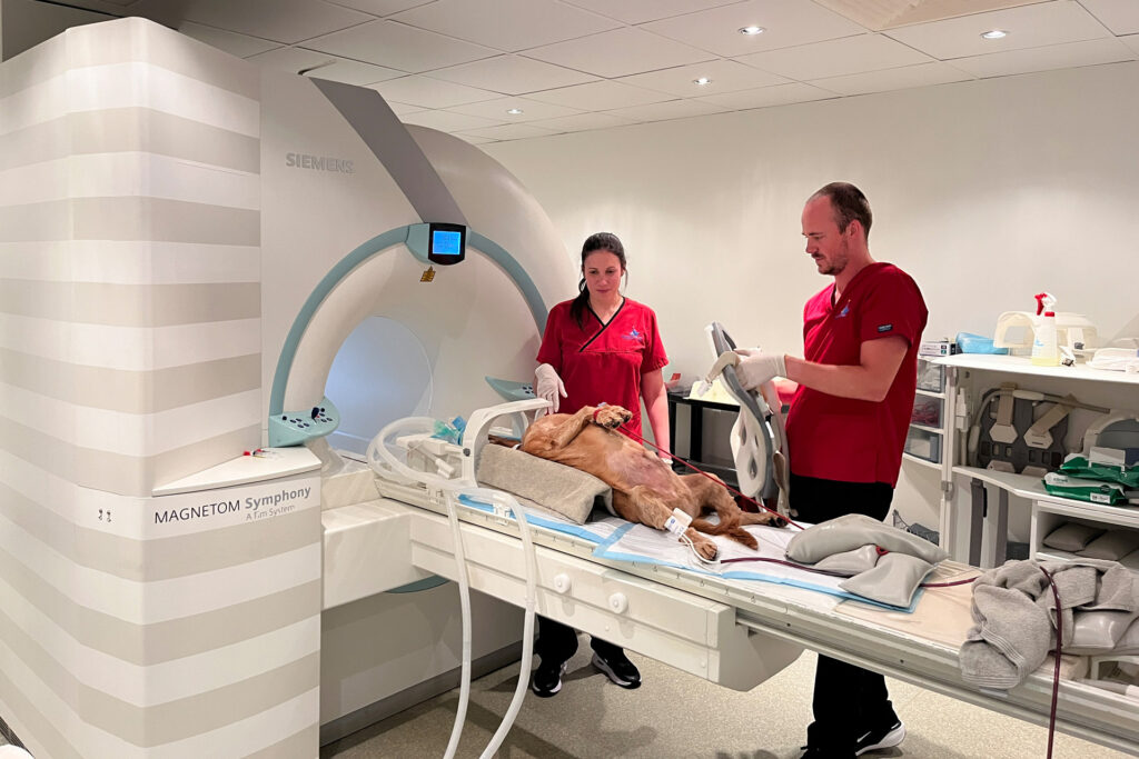
Using strong magnetic fields and radiofrequency energy, MRI (magnetic resonance imaging) produces highly detailed images of the body’s soft tissues. Image acquisition takes longer, but MRI is vital for assessing the brain and the spine as well as joint structures such as ligaments and tendons.
Different imaging techniques offer different perspectives.
Some common examinations:
- MRI of the brain
- MRI of the spine
- MRI of the shoulders
- Some of the questions it can help us investigate:
- Has the patient suffered an intervertebral disc extrusion?
- Is there degeneration of the lumbosacral joint and associated nerve compression?
- What might be causing ongoing seizures?
Images behind the investigations
Just a few examples of the areas we can examine using advanced diagnostic imaging (L – R):
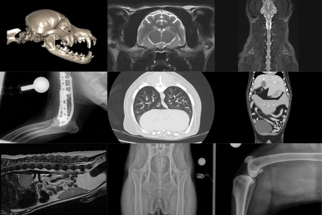
- Teeth
- Brain
- Cervical spine
- Elbows
- Lungs
- Abdomen
- Lumbar spine
- Hips
- Stifles
More than just taking a picture
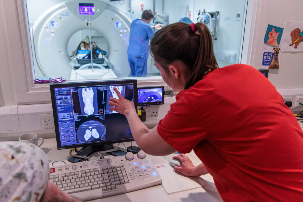
Every image has a purpose – behind each imaging examination there is a clinical question, and our radiography team works closely with our clinicians to help obtain the information needed to investigate and manage each patient’s condition.
Medical imaging can help:
- Investigate symptoms – acute and chronic
- Identify injuries and abnormalities
- Support or rule out potential diagnoses
- Guide treatment decisions and surgical planning
- Aid custom implant planning and engineering
- Assess implant positioning
- Monitor certain conditions over time
Imaging is an important and integral part of the patient journey, helping our team make informed decisions about patient care.
A special thank you to our wonderful radiography team for the expertise, dedication and care you bring to your work every day.