What is patellar luxation?
The patella (kneecap) is a small bone that sits within the tendon of the quadriceps muscle (large thigh muscle) – the tendon crosses the knee in the front and attaches on the tuberosity (bony bump) of the tibia (shin bone).
The patella acts as a fulcrum during extension of the knee; it glides up and down within a groove on the front of the femur (thigh bone), which is part of the knee. In some dogs, the patella luxates (dislocates) out of this groove. The consequence of this luxation is an inability to properly extend the knee and when this mechanical dysfunction is chronic, varying degrees of pain and osteoarthritis can also be part of the lameness.
How can I tell if my pet has patellar luxation?
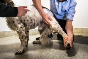
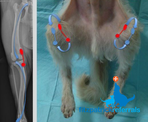
Patellar luxation is a common condition that mostly affects dogs, although cats can occasionally be affected. It is more common in smaller dogs, but medium and large dogs can also be diagnosed. Dogs with a “bow-legged” stance are particularly predisposed.
The age at onset of clinical signs is variable: most dogs already start to show signs as puppies or young adults; although first clinical signs can also be detected later in life in adult dogs. Often, a characteristic “skipping” lameness is noticed, where dogs will limp for a few steps and then quickly return to a normal gait. Some dogs will limp continuously and if both knees are affected, they will have a stiff, awkward gait with knees that do not extend properly.
What is the cause of patellar luxation?
The condition is primarily of genetic origin and the consequence of the selective breeding of dogs with a preferred (bow-legged) conformation. Animals are born with normal knees, but begin to develop abnormalities of the bones and muscles of the hind limbs early in life.
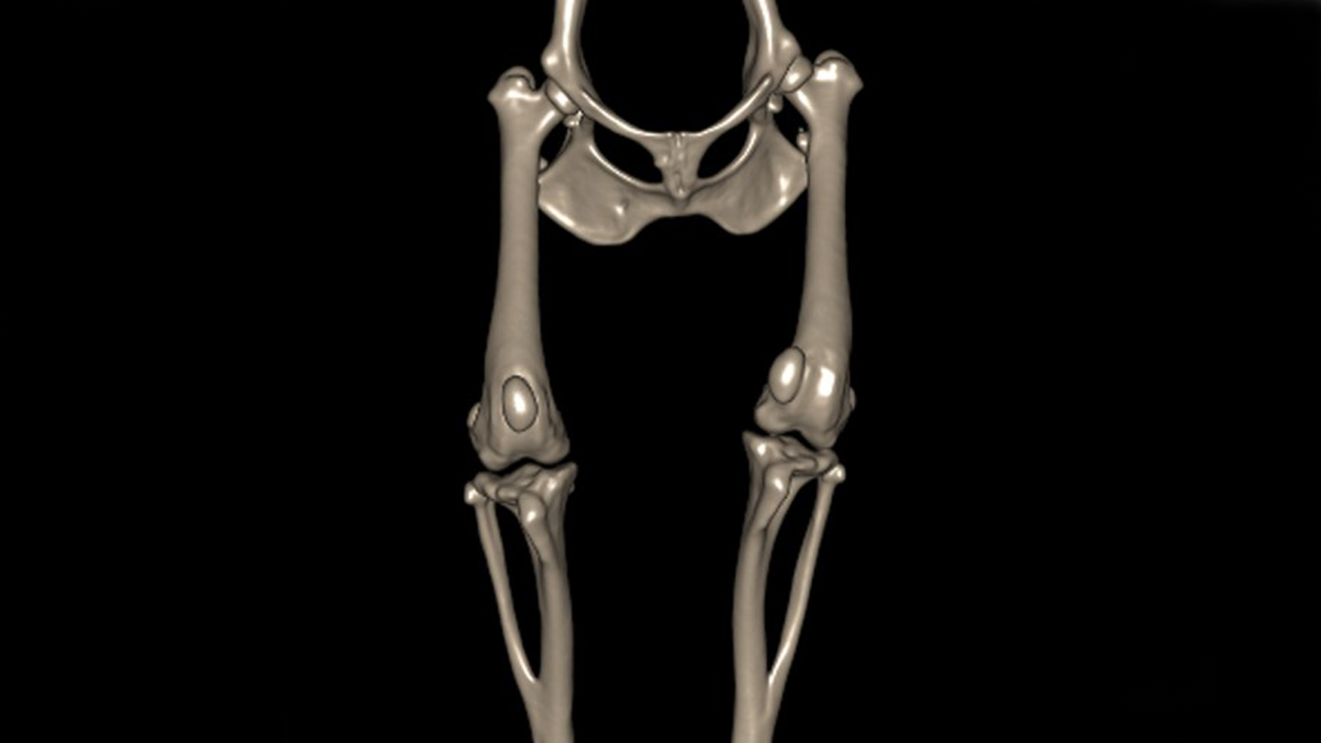
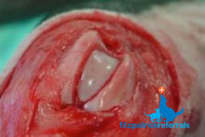
Patellar luxation results from malalignment of the so-called “quadriceps extensor mechanism”, which consists of the quadriceps muscle, the patella with the patellar tendon, and the tibial tuberosity, which together extend the knee. The most common direction of luxation is to the medial aspect (inside) of the knee. When the powerful quadriceps mechanism starts to displace in this direction during the dog’s growth, it acts as a bowstring and causes the femur (thigh) and tibia (shin) to deform into a pronounced outwards bow. Subsequently, the groove in which the patella slides during knee extension does not develop properly and becomes shallow; the resulting limb deformities become self-perpetuating at this stage (see photograph above). Patellar luxation is usually one aspect of a complex limb deformity, that can vary in severity. Patellar luxation to the lateral aspect (outside) of the knee is less common and typically occurs in large dogs. Traumatic patellar luxation is rare.
What is happening inside an affected joint?
As the patella slides in the shallow groove, it often slides on the groove’s ridges when it luxates and can wear off the cartilage of the patella itself and of the ridges. This causes pain and triggers a cascade of joint inflammation, resulting in progressive osteoarthritis. In addition, the abnormal pull of the quadriceps mechanism causes internal rotation of the tibia relative to the femur that can strain other structures within the knee, including the cranial cruciate ligament (CCL). The longer the patella spends outside its normal groove, the shallower the groove becomes, the easier and more often the patella luxates and the more cartilage gets damaged.
How is patellar luxation diagnosed?
Patellar luxation is typically diagnosed following a multimodal evaluation process between you, your primary care vet and a specialist orthopaedic surgeon.
Patellar luxation is usually picked up during clinical examination by your primary care vet at a routine health check, or when you have highlighted to your primary care vet that your dog shows an abnormal “skipping” gait during exercise. Usually, upon diagnosis of a luxating patella, your primary care vet will refer your dog for assessment by a specialist orthopaedic surgeon.
During your initial consultation at Fitzpatrick Referrals your dog will be examined by one of our orthopaedic clinicians. Your dog will then be admitted to the hospital to take radiographs (x-rays) of the affected knee under sedation or a short general anaesthesia. Your dog may also require additional diagnostic imaging such as a CT scan, which will be performed by our advanced diagnostic imaging team. Your dog will receive one-to-one nursing care throughout the process by one of our nurses from the prep nursing team who are all highly trained and experienced in sedation and anaesthesia. When patellar luxation is diagnosed after combining the orthopaedic exam results and diagnostic imaging findings, your dog may require corrective surgery.
What is the grading system for patellar luxation?
The grading system for patellar luxation is based on how mobile the patella is relative to the groove at the base of the femur.
- Grade 1: The patella can be luxated with manual pressure but is otherwise stable within the groove.
- Grade 2: The patella spontaneously luxates and reduces (goes back) into its normal position in the groove during knee motion; this is typically associated with a skipping lameness.
- Grade 3: The patella is permanently luxated but can be manually replaced into the groove.
- Grade 4: The patella is permanently luxated and cannot be manually replaced into the groove.
How is patellar luxation treated?
Occasionally, patellar luxation is an incidental finding during a routine physical examination. In adult dogs, non-surgical treatment might be the best option in this case. In immature animals, surgical management may be more appropriate in order to try to prevent the development of severe limb deformities as they grow; the timing of surgery will be decided on a case-by-case basis.
In general, surgery is strongly advised early in the course of the disease for grade 3 and 4 patellar luxation to mitigate progressive limb deformities and osteoarthritis. For grade 2 medial patellar luxation, surgery is usually recommended only for dogs exhibiting significant clinical signs, such as lameness. These dogs should have regular re-evaluations to determine the best timing for surgery. Surgery is never advised for grade 1 patellar luxation.
Could delaying treatment do more damage?
Patellar luxation, when severe, can cause skeletal abnormalities such as increased bowing of the femur or tibia. In milder cases, we often see cartilage loss from the underside of the patella and/or the surface of the groove as the cartilage is repeatedly shaved off from the patella when it luxates out of the groove.
Dogs with medial patellar luxation – when the patella luxates to the inside of the knee – are also more prone to developing cranial cruciate ligament disease. As with most diseases, if the luxation is causing a clinical problem, then the earlier it is addressed the better it is for the dog, in the short and the longer term.
Non-surgical treatments for patellar luxation
The cornerstones of non-surgical treatment for patellar luxation are body weight management, physiotherapy, exercise modification and medication (anti-inflammatory painkillers). These same tools are also important in the short-term management of dogs who have surgery for patellar luxation. However, the primary goal of surgery is to minimise the requirement for long-term exercise restriction and medication.
At Fitzpatrick Referrals, we are able to provide you with a rehabilitation plan for your dog if conservative management of patellar luxation is indicated. This is delivered through our dedicated rehabilitation service, chartered made up of experienced chartered physiotherapists and hydrotherapists. Your orthopaedic clinician will coordinate an appointment with one of our chartered physiotherapists, who will carry out a thorough clinical examination. Following this, an individual rehabilitation plan will be created for your dog, including a home exercise plan for you to follow. Most appointments are carried out on an out-patient basis and your chartered physiotherapist will regularly evaluate your dog’s progress and amend your home exercise plan as necessary.
Surgical treatments for patellar luxation
Surgical treatments are recommended for dogs with intermittent or permanent lameness as a result of patellar luxation. Many surgical techniques exist and the primary goal is to restore normal alignment of the quadriceps muscle mechanism relative to the entire limb. This often requires reshaping of the bones – tibia and/or femur – in addition to reconstruction of the soft tissues around the knee.
Tibial tuberosity transposition
The most important component of the corrective surgery is to realign the path of the tendon that connects the patella to the tibia. This is done by changing where the tendon attaches. Because bone heals better than tendons, the tibial tuberosity (small piece of bone where the tendon attaches) is cut and moved to a better position. It is fixed into place with small pins and the bone usually heals over the following 4-8 weeks. Often wire is placed in addition to the pins, to counteract the pull of the quadriceps muscle. This helps restore the quadriceps mechanism that extends the knee and improves how the patella moves.
Femoral varus osteotomy
Femoral varus osteotomy is most commonly performed on larger dogs and dogs with higher grades of patellar luxation that results from a severe bow in the lower segment of the femur – this segment is straightened with the surgery. The bone is cut – sometimes in three dimensions – and a wedge of bone is taken out, the gap is closed by realigning the bone segments and the bone is repaired with a plate and screws. Performing a CT scan before surgery is particularly important when planning such corrections, as it gives a 3D orientation of the bone cut that needs to be made. One such cut is a so-called “dome osteotomy”, which allows re-angulation of the bone segments in any direction.
Recession sulcoplasty
When the groove in which the patella slides in is very shallow, the groove can be deepened to give the patella a better seating. A wedge or block of bone including the cartilage is cut out and repositioned into the created bone gap in reverse. Often, when deep seating is required, bone is rasped or filed off the bone block or wedge; block-shaped deepening of the groove is usually superior to wedge-shaped deepening.
Soft tissue reconstruction
In most dogs, the soft tissues on the inside of the knee are too tight and soft tissues on the outside of the knee are too loose. Therefore, the soft tissues on the inside are often released and on the outside are tightened to reestablish soft tissue balance around the knee.
Trochlear ridge replacement (TroRR)
The patella slides within the trochlear groove of the femur; this groove is bordered by the medial (inside) trochlear ridge and the lateral (outside) trochlear ridge. In dogs with severe limb deformities, the trochlear groove is usually too shallow. In particular, the medial ridge is too low so that the patella rides over the ridge and luxates out of the groove. We have designed a custom-made implant, based on patient-specific CT scans taken before surgery, that replaces the medial trochlear ridge; this results in a tall prosthetic ridge that prevents the patella luxating medially.
Will my dog be able to exercise normally after patellar luxation surgery?
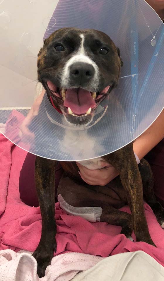
Your dog will initially have to undergo a standard period of 6-week exercise restriction, usually involving cage rest combined with a limited lead exercise programme prescribed by your orthopaedic surgeon.
At Fitzpatrick Referrals, we will provide you with mobility slings to prevent your dog from struggling or falling when walking after surgery. In addition, you can get further advice on physiotherapy and hydrotherapy options that are provided on an outpatient basis through our rehabilitation service. During your initial rehabilitation appointment, one of our chartered physiotherapists will assess your dog, design a patient-specific rehabilitation programme and advise you on how often rehabilitation appointments are required.
Following a 6-week postop reassessment with your orthopaedic surgeon, and providing your dog is making good progress, there is no reason why they cannot enjoy the same full range of exercise as before surgery.