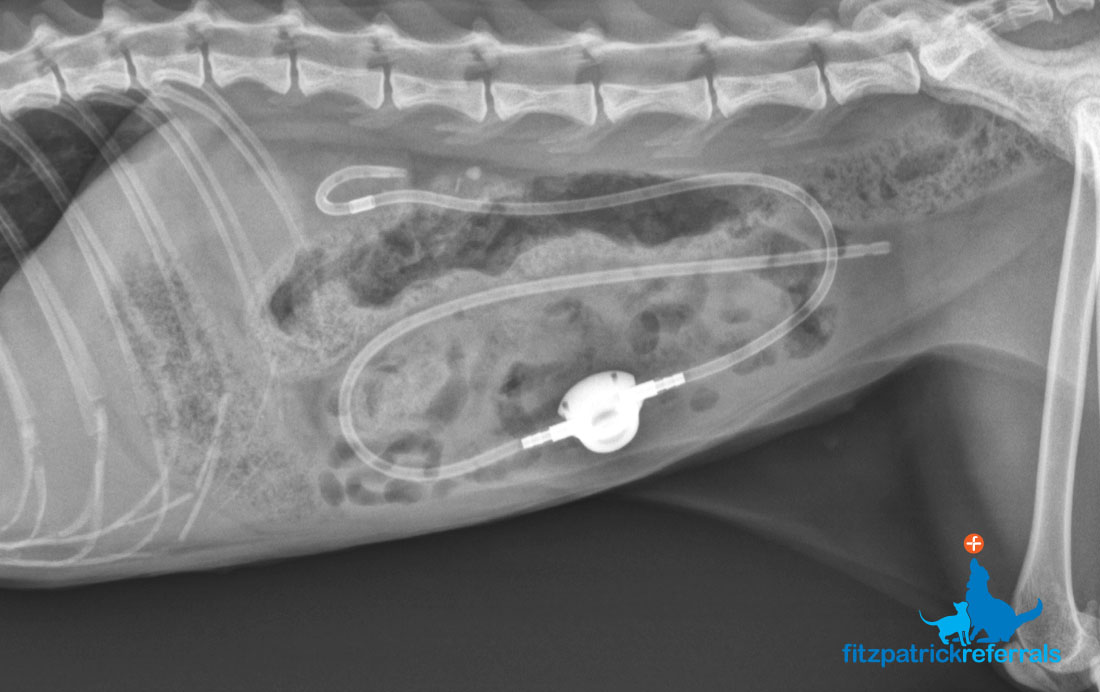
Subcutaneous Ureteral Bypass (SUB)
A subcutaneous ureteral bypass (SUB) device is used to treat obstructed ureters (the ureter is the tube carrying urine produced by the kidneys to the bladder). Ureters most commonly block due to stones but can also obstruct due to tumours, strictures (scars) and blood clots. A SUB device involves tubing being placed into the kidney and then into the bladder via a port, which is located under the patient’s skin. It therefore bypasses the obstruction and effectively provides the patient with a new ureter. Ureteral obstruction due to stones is more common in cats than dogs and it is recommend that any patient with increased kidney markers on blood analysis undergoes an ultrasound to assess the kidney for potential obstruction.
Why do animals get ureteral obstructions?
Most obstructions of the ureters are due to stones which usually contain calcium. Certain breeds are predisposed to calcium stone formation and certain medical conditions, including cancer and endocrine diseases, can result in increased blood calcium which leads to stone formation. Cats can also have high blood calcium levels which predisposes to stone formation.
How is a SUB placed?
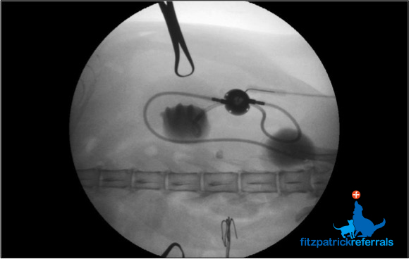
SUBs are placed during an open abdominal surgery using fluoroscopy (live video x-ray). The SUB port is placed below the patients’ skin. After the SUB is placed most animals remain hospitalised for 72 hours to allow ongoing intravenous fluid therapy and pain relief.
What are the benefits of a SUB?
Traditional surgery on a ureter is associated with a 20% death rate due to leakage of urine at the surgical site. Since ureters in veterinary patients are so small (1mm in a cat) there is also a risk of stricture (scar) formation at the site of the stone removal and this occurs in up to 40% of patients. Since the SUB device placement does not involve touching the ureter surgically the previously mentioned risks do not exist. The SUB device is associated with a 5% death rate, which compares very favourably to traditional surgery.
What are possible complications seen with SUBs?
Reported complications include blockage of the SUB with a stone, recurrent urinary tract infections and kinking of the SUB tubes. Blockage of the SUB device with stones is reported in 25% of cases but only 12% (roughly 1 in 8 cases) would require a second procedure to unblock the device. Most of the complications reported following SUB placement can be prevented by ensuring the procedure is carried out by a veterinary surgeon who has trained in Interventional Radiology.
What monitoring is required after SUB placement?
We recommend the SUB device is flushed via the port one month after placement and then every 3-6 months for life. This would normally be performed at a referral practice. The flushing is normally done without any sedation or anaesthesia. At the same time, a sample of urine will be taken from the device to ensure no infection has developed and blood work will be checked to monitor kidney function.

