Three-year-old Foxy was first referred to our orthopaedic service last summer (2025) for investigations into her waxing and waning left forelimb lameness, which was limiting her ability to exercise.
Diagnosis
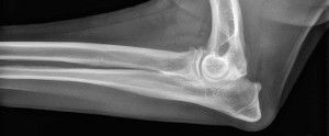
CT scans taken confirmed she has a condition called fragmented medial coronoid process, which is when a piece of bone on the inside of the elbow joint has cracked, chipped or broken off, which has the effect of rubbing against the other bones like a pebble in a shoe, causing pain, swelling and arthritis.
Surgery
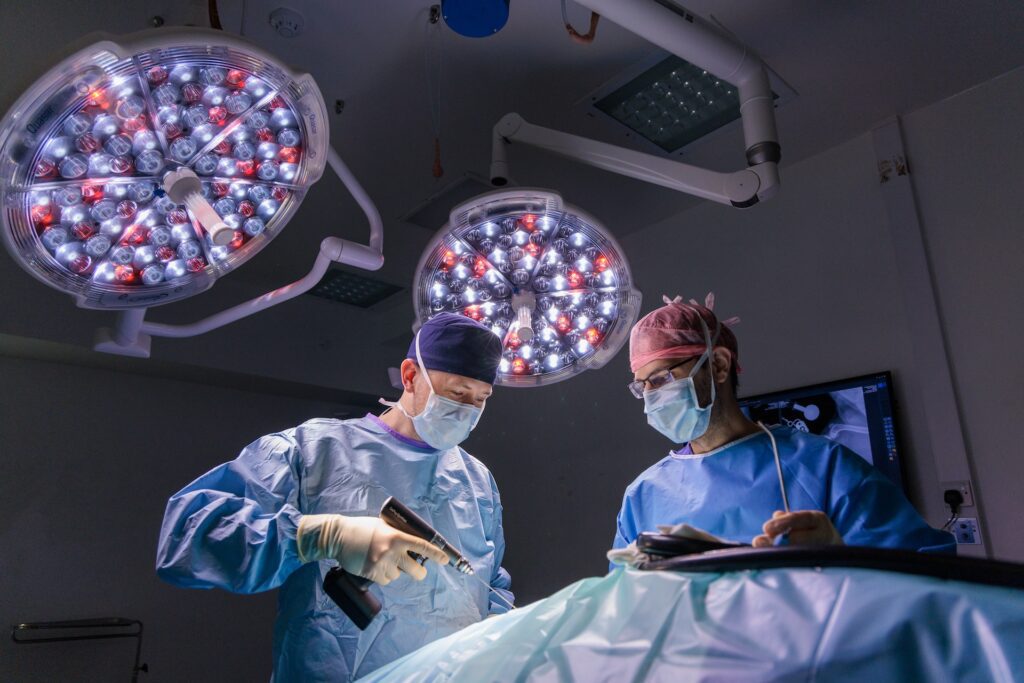
Foxy underwent a bilateral elbow arthroscopy, which is a minimally invasive surgical procedure looking at both joints using a tiny camera that allowed her surgeon to remove loose bone chips and damaged cartilage. In addition, she also had a biceps ulnar release procedure (BURP) performed on both elbows. Whilst under the same general anaesthetic, Foxy also had a small sample of fat tissue taken, which was processed in our in-house laboratory to grow adipose derived mesenchymal stem cells (AdMSCs).
Recovery
Following her surgery, Foxy was discharged with a home physiotherapy exercise plan. Her recovery involved six weeks of crate rest, with short lead walks only.
Stem cell therapy
Six weeks later at her recheck, Foxy showed a satisfactory recovery from surgery. In dogs with this condition, mild persistent or recurrent lameness is common due to the nature of the condition. However, for continued management of her osteoarthritis, she had both elbow joints injected with adipose derived stem cells and platelet-rich plasma (PRP).
A few months later, we were pleased to receive this update from Foxy’s family:
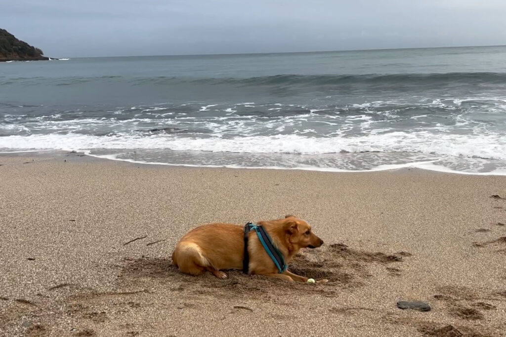
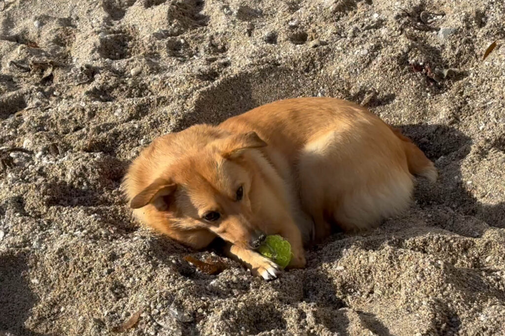

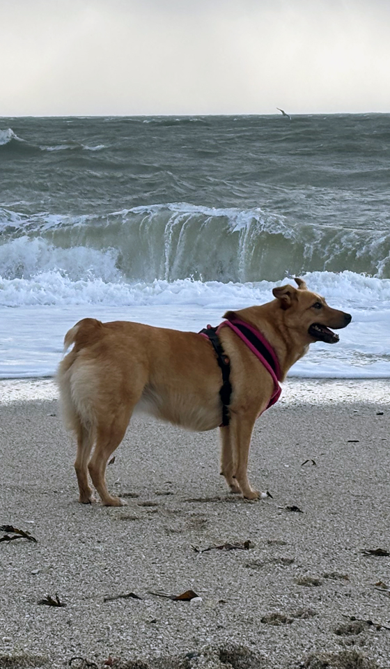
Foxy returned to us 7 months later for her second injection of stem cells and PRP, and continues to live life to the full enjoying the beautiful Cornish beaches.
Thank you to Cornish Paws Vets for your referral.
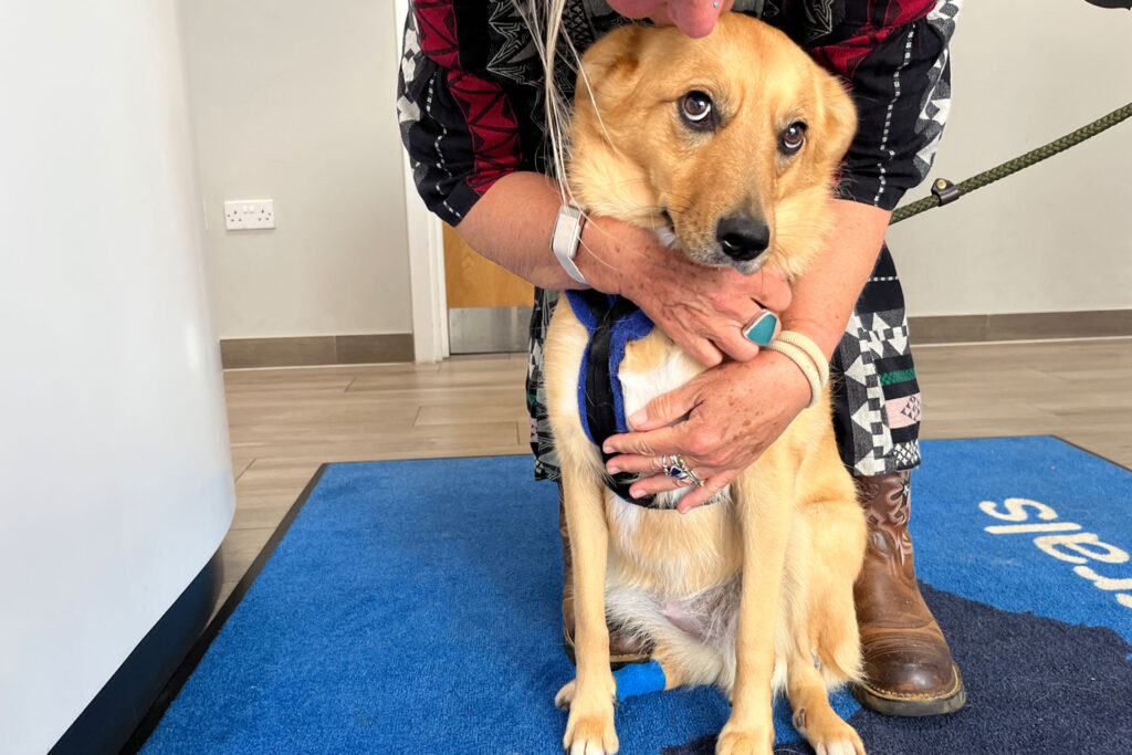
Learn more about developmental elbow disease (elbow dysplasia) and stem cell therapy
Services used in this story

Fitzpatrick Referrals is dedicated to the prevention and treatment of orthopaedic, neurological and dental diseases in small animals.